
Shengtao Yu Wins Award for “Fiber-Array-to-Chip” Paper
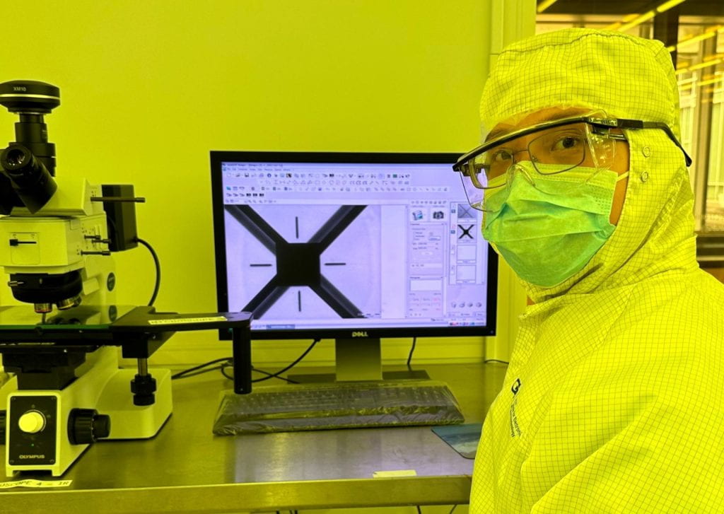
Ph.D. student Shengtao Yu has won the Intel Outstanding Student Paper Award for his paper “Scalable Fiber-Array-to-Chip Interconnections with Sub-Micron Alignment Accuracy” [1] presented at the Electronic Components and Technology Conference (ECTC). The results of this research on co-packaged optics (CPO) will support next generation processing systems which will require optical connectivity in very close proximity to the electronics. This technology being developed enables arrays of optical fibers to be connected directly to the semiconductor chips that do the data processing.
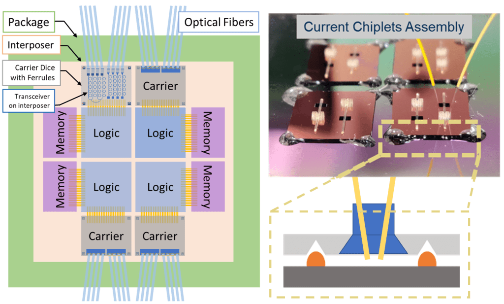
This research includes innovative design and technology to enable the passive alignment and interconnection of an array of optical fibers to an underlying photonic integrated circuit (PIC). The key to the demonstrated technology is a passive silicon chiplet, which contains 3D printed slots for fiber insertion, that precisely self-aligns to an underlying PIC at sub-micron scale. This has the potential to eliminate active alignment techniques commonly used in photonics packaging and enable a scalable and silicon-based solution to fiber alignment and coupling. Photonic interconnection between modern 2.5D packages with PICs and optical fibers is a current key bottleneck. This work will potentially lead to improved and scalable CPO architectures, enabling massive off-package bandwidth and reduced energy consumption of interconnects in data centers, which will be important to meet the rapidly increasing packaging demands of artificial intelligence applications and beyond.
[1] S. Yu, T. K. Gaylord, and M. S. Bakir, “Highly scalable fiber-array-to-chip interconnections with sub-micron alignment accuracy,” in IEEE Electronic Components and Technology Conference (ECTC), 2023, pp. 748-752.
Source: IEEEXplore
Optics Laboratory Research Receives “Spotlight on Optics” Recognition
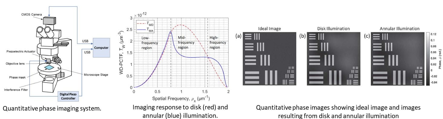
The recent research of Pranav Kulkarni, Yujin Bao, and Tom Gaylord has been selected by Optica (formerly Optical Society of America) for “Spotlight on Optics” recognition. Centered on the ECE Master’s thesis research of Pranav Kulkarni, the resulting Applied Optics paper “Annular illumination in 2D quantitative phase imaging: a systematic evaluation” [1] has been highlighted by Optica to receive this recognition. According to Optica “Only two papers are highlighted from our respective journals each month from among the scores of fine articles published.”
Quantitative phase imaging (QPI) is having a significant impact in biomedical research revealing heretofore invisible details of cellular composition and function. Early onset detection of numerous diseases including breast cancer and prostate cancer has resulted from the application of QPI. More such breakthroughs are anticipated in the near future. A critical review of the Kulkarni, Bao, Gaylord paper for “Spotlight on Optics” by Prof. David Paganin of Monash University in Australia describes this ECE research: “Quantitative phase imaging (QPI) takes another step forward with this interesting study on the influence of annular illumination. Understood in its broadest sense, QPI has made important contributions to optics over many decades, for a variety of radiation and matter-wave fields. This family includes visible light, electrons, x-rays and neutrons. The current paper, by Kulkarni and coauthors, studies annular illumination in two-dimensional QPI for visible-light microscopy. Particular attention is paid to the comparison between annular illumination and disk illumination. Defocus is the method of choice for this study. In particular, defocus generates the phase contrast that is subsequently decoded to achieve QPI. As is the case with Gabor’s immensely fruitful concept of inline holography, the QPI imaging protocol is a two-step process. First, multiple defocused images are measured, that contain coded phase information. Second, the measured focal series of images is decoded, to provide quantitative phase information. Stated differently, this is phase-retrieval computational imaging. Optical software constitutes an intrinsic part of the QPI imaging system, whose optical components are therefore both actual and virtual. The work is important and interesting. I warmly recommend it to your attention.”
[1] P. P. Kulkarni, Y. Bao, and T. K. Gaylord, “Annular illumination in 2D quantitative phase imaging: a systematic evaluation,” Appl. Opt., vol. 61, pp. 3409-3418, Apr. 20, 2022.
Source: Optica.org
Optics Laboratory Researcher Vishrutha Arun Goes to Columbia Medical School

Vishrutha Arun came to Georgia Tech with an interest in its Pre-Med program. Supported by the prestigious Zell Miller Scholarship, she entered the Scheller College of Business and concentrated on information technology management with, of course, a minor in biology.
As she progressed in her program, she wanted to add a meaningful research component to her background. As a result, she joined the Optics Laboratory research team in ECE that was doing research on fiber optic gratings. Vishrutha added a new dimension to this research by investigating medical applications of these devices. Sensors for detecting antigens and antibodies was a major application. However, haptic (or sense of feel) applications were particularly interesting as these had important clinical, surgical, and dental applications. Her undergraduate research work in the Optics Laboratory has led to new directions of research that utilize the quantitative phase imaging capabilities developed in the Optics Laboratory.
When it came time to apply to medical schools, Vishrutha decided to apply to schools with a strong dental program as well as with a significant research emphasis. There are always applications to multiple schools and with pros and cons to consider. However, all things considered, Columbia University was the top-rated school for her interest and her top choice. Her strong record, including research, carried the day and she was admitted to Columbia. Vishrutha has found it to be perhaps more rigorous than expected. Currently she is taking classes that cover biochemistry, immunology, pharmacology, genetics, histology, pathology, and anatomy. She is in the thick of it. We take this opportunity to thank her for her research contribution in the Optics Laboratory and to wish her all the best going forward
GTRI Best Paper Award
The Georgia Tech Research Institute (GTRI) has selected the lidar paper G. B. Popko, Y. Bao, T. K. Gaylord, and C. R. Valenta, “Beam path intersections between two coplanar lidar scanners,” Opt. Engr., vol. 58, p. 033103 (14 pp.), Mar. 2019 to receive the GTRI Best paper award for 2019.
Motivated by the increasing use of lidar systems in semi-autonomous and autonomous vehicles, this seminal paper presents a mathematical model describing when two circularly scanning lidars can have direct and indirect interference between the scanners. Such interference may result in erroneous range data.
The fraction of time the scanners’ beams intersect is analyzed as a function of rotational frequencies and their reference phases. It is shown the minima and maxima of this fraction are 0 and 1/2 with most configurations resulting near 1/4.

A procedure to adjust the scan rates and phases to minimize this intersection time (including zero intersection) thus reducing the risk of mutual interference is also outlined. Four configurations that produce zero intersection between two scanners are presented. Configurations of three and four scanners are also presented for which no intersections between the scanners’ transmission paths occurs. This best paper award was announced in June 2020.
Source: SPIE Digital Library
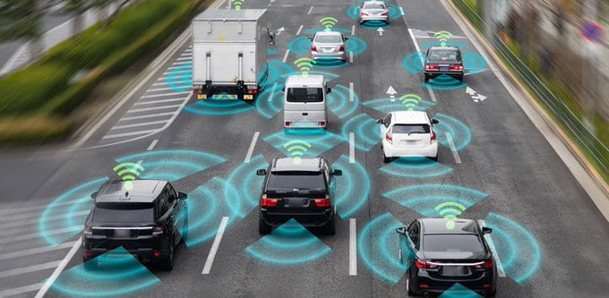
Lidar Drives Toward the Future
Researchers’ new methodology confirms model of lidar interference
The future of automobiles is autonomous. Advances in light detecting and ranging (lidar) sensors and other sensing technologies are quickly bringing that future into the present. However, the assumption that self-driving vehicles will result in safer roads and highways by reducing human error relies on the idea that sensing technologies will advance enough to permeate the automotive market on a large scale. Technologies such as millimeter wave radar, ultrasonic sensors, and chromatic imaging have already done that, but development of lidar for self-driving cars has lagged, partly because little research has been done into the risks of signal interference among different vehicles’ lidar systems. Research has shown that interference between lidars can happen when the beam sent from one lidar is scattered and returned or transmitted directly to another lidar. Lidar interference has been shown to happen if the lidars’ optical axes intersect and if a hard or volumetrically scattering target is present where they intersect. If multiple self-driving vehicles are at a busy intersection, the lidars from each vehicle would have the potential to interfere with the others. It is possible that incorrect data could be returned because of the presence of other lidar sources, possibly resulting in collisions.
Mathematical Model
Given the pressing need for lidar technology to adapt for use in autonomous vehicles, mathematical models may seem like an overly academic consideration. However, models predicting the risk of interlidar interference are rare in scientific literature, and the existing works do not offer a quantitative look at the interference mechanisms. The few works that do exist have shown that interference can be modeled with simple geometry. Mathematical considerations are necessary now to direct engineering requirements, with simultaneous consideration of safety and deployment at scale.
Gerald Popko and colleagues, reporting in Optical Engineering, presented a methodology to analyze interference effects between spindle-type lidar scanners that would likely be used in autonomous vehicles and would be suited for general multilidar applications. Their experiment used the methodology to analyze conditions for interference between lidars. They tested four different configurations using multiple lidars. Their tests confirmed previous research into interference and established standard reference points for a geometric lidar model. The results supported at least two forms of interference that had previously only been theorized: direct interference and scattered interference.
While direct interference was found to occur more frequently, scattered interference resulted in much larger range error. In the case of self-driving vehicles, scattered interference could present more risk because it could create larger errors than direct interference would.
The authors believe that better understanding lidar interference can continue to be developed using the models proposed in previous research along with their enhanced characterization of lidar interference modes. “Though simple geometry was used to develop an initial understanding of the phenomenon, the analysis methodology can be applied to more complex situations such as out-of-plane scanners, complex target geometries, non-Lambertian surfaces, and multiple reflection environments. With this collective understanding, lidar engineering can account for this phenomenon through quantitative and qualitative anticipation of its effects to ensure the safe and reliable application of lidar sensors in greater density.”
Source: SPIE Digital Library
Optics Laboratory Alumna Ashton Hattori Now at MIT

As an undergraduate student in electrical and computer engineering at Georgia Tech, Ashton Hattori seemed to excel in everything that she did. In addition to being a top student and taking a full academic load, Ashton found time for research and volunteer work. She was an Opportunity Research Scholar (ORS) in the Optics Laboratory where she implemented label-free quantitative phase imaging for immobilized biological cells in glass microcapillaries for applications in bioinformatics and early cancer detection. This research resulted in an overall second place research award in the campus-wide ORS program. Ashton spent a summer as an intern at the University of California, Berkeley working on energy-efficient optical interconnects on III-V semiconductor chips. During her program at Georgia Tech, she volunteered by guiding and mentoring at-risk children after school by helping them develop basic skills in the setting of bicycle maintenance and repair. Ashton graduated with highest honors and has now gone on to become a PhD student in electrical engineering and computer science at the Massachusetts Institute of Technology (MIT). There, she is doing research in the Photonics and Electronics Research Group. Ashton is collaborating with a team at Lincoln Laboratory to develop CMOS-compatible silicon photonic chips for laser cooling of trapped ion quantum computing systems. This challenging work seeks to integrate a 422nm-operating-wavelength system with CMOS-compatible fabrication processes. This is difficult, but Ashton has a history of accomplishing difficult tasks!
ECE Experimental Research During the Pandemic
To respond to the escalating COVID-19 pandemic and to keep faculty, staff, and students safe, Georgia Tech implemented a “Research Ramp Down Plan” that closed all on-campus research laboratories on March 19. “Work-from-home” became the necessary mode of operation. Theoretical and computer-based research have adjusted to the shutdown without major difficulties. However, experimental laboratory-based research largely came to a halt. This unfortunate situation will likely cause delays in student progress toward graduation, delays in deliverables to sponsors, delays in getting data for scheduled presentations, and more.
In this setting, clever ECE students are coming up with “work-around” ways of continuing their experimental research! An example of this is the ongoing research being conducted by Ji Ye (JC) Chun. Ms. Chun is a fourth-year senior who is transitioning into the Master’s degree program thesis option. She is one of our perfect 4.0 graduates and is supported by a PURA award. Her research in the Optics Laboratory involves quantitative phase imaging of biological cells and fiber optic devices. This overall area of work is supported by a new National Science Foundation grant to Prof. Tom Gaylord to investigate the highest possible resolution (in space and in refractive index) that can be achieved in imaging of phase objects such as optical fibers and living cells. A crucial phase of this research is the preparation of the samples for microscopic imaging. With the disruption of on-campus research, JC has moved a few small pieces of equipment and some critical materials to her nearby apartment and set up a “work-from-home” laboratory.
However, she still needed a microscope to continue her research. Multi-thousand-dollar microscope systems are available in the Optics Laboratory. But these are not available in her apartment. Amazingly, JC located a very small portable microscope (50X to 1000X) with bright adjustable LED illumination that also incorporates a digital wireless connection. This was purchased with NSF funds. The cost including shipping was less than $50! In this case, the virus crisis has led to some unanticipated increased capability! This has enabled JC to continue her sample preparation research in her apartment.
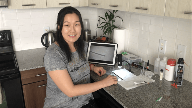
She is assisted by Lucky, her faithful dog. Lucky gets excited every time JC starts working in her kitchen laboratory, thinking that she is preparing food. But alas, JC is only developing a protocol for sample preparation, not making a tasty snack to be shared. The innovative approaches of our students are overcoming some of the pandemic setbacks. Our hats are off to JC for her dedication to research during these challenging times and for setting an example for others to emulate.
Source: Georgia Tech